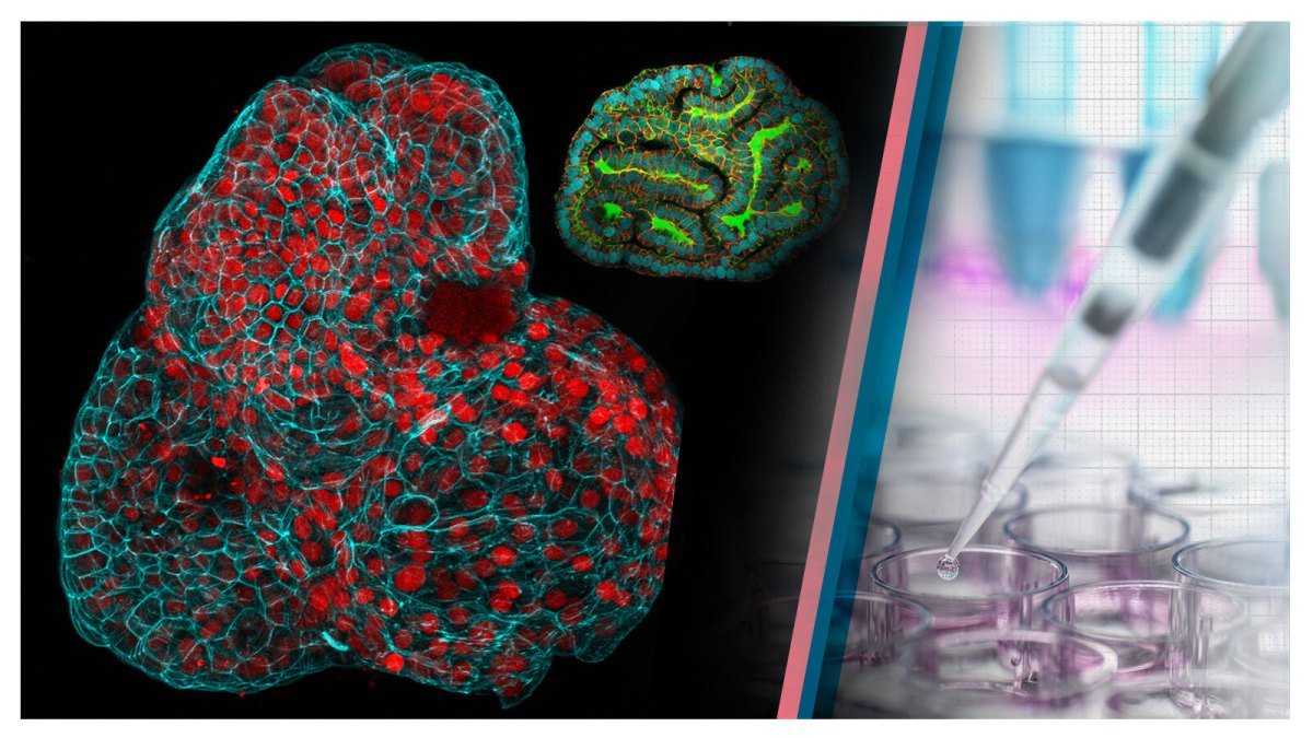
Researchers from University College London and Great Ormond Street Hospital have developed a technique to grow miniorgans, or “organoids,” from cells collected from amniotic fluid samples taken during active pregnancies. This marks the first time miniorgans have been grown from cells during ongoing pregnancies. Miniorgans, simplified structures resembling real organs, are used for testing new medical treatments and studying organ functions. The scientists envision this approach aiding in monitoring and treating congenital conditions before birth and developing personalized therapies for babies in the womb. The technique also bypasses regulatory challenges associated with obtaining stem cells directly from fetal tissue.
Key Points:
- Researchers collected cells from amniotic fluid samples taken during 12 pregnancies and grew miniorgans from cells taken during active pregnancies.
- Miniorgans, or organoids, are tiny structures used to test medical treatments and study organ functions.
- Previously, miniorgans were derived from adult stem cells or fetal tissue after an abortion. Growing them from amniotic fluid cells overcomes regulatory challenges related to fetal tissue.
- The tissue-specific stem cells were shed by the fetus, allowing identification of cells from the lungs, kidneys, and intestines.
- This approach could help monitor and treat congenital conditions before birth, providing personalized therapies.
- Researchers worked with colleagues in Belgium to study the development of babies with congenital diaphragmatic hernia, growing lung organoids from affected foetuses before and after treatment.
- The ability to study functioning prenatal miniorgans is seen as a step toward more detailed prognoses and effective treatments.
- While still in the early stages, the research holds promise for advancing prenatal medicine.